Histology
The OBRL offers a wide range of histology services including paraffin (soft tissue, decalcified bone), plastic (undecalcified bone, implants), and frozen embedding methods, as well as imaging and analysis. We can complete high resolution digital imaging and static and dynamic histomorphometery analysis in house and routinely work closely with a board certified veterinary pathologist. The OBRL routinely works with investigators through every stage of a project from study design to tissue handling and final outcomes.
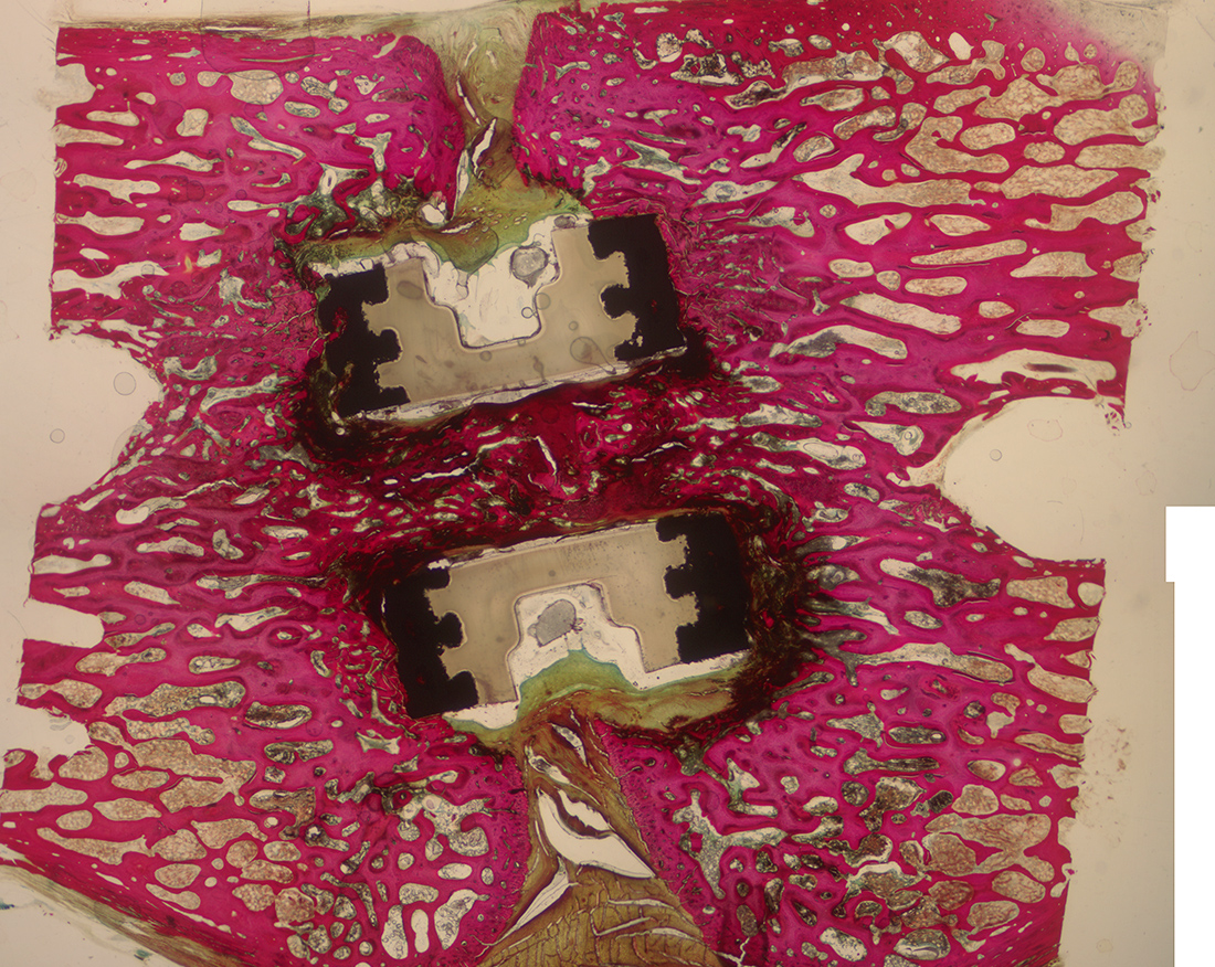
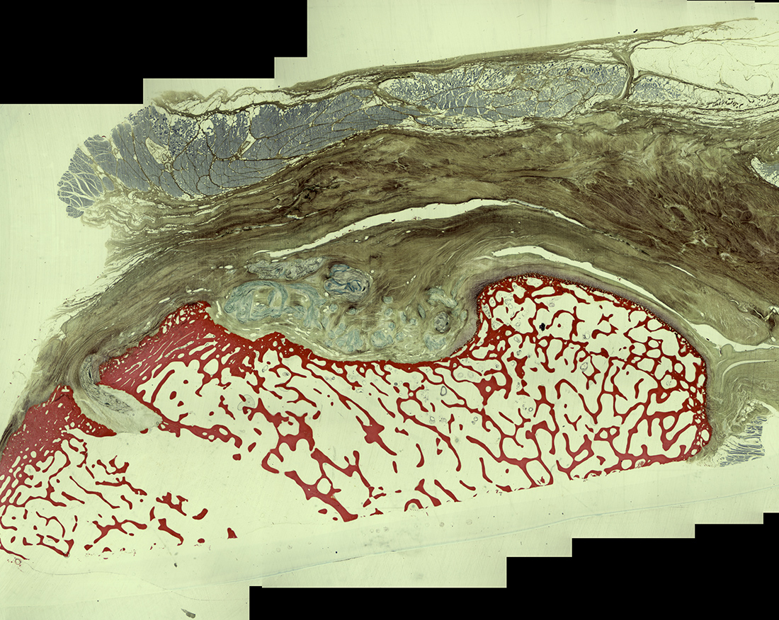
Hard Tissue Processing
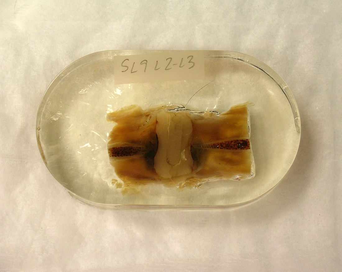
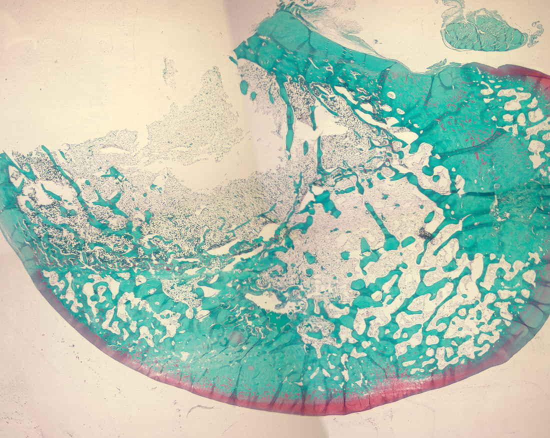
Hard tissue histology techniques are used for samples containing undecalcified bone, hard implants (plastic, metal, ceramic) or fluorescent markers. Methylmethacrylate is used to embed samples, which are then cut and ground using the Exakt method or sectioned on a rotary microtome.
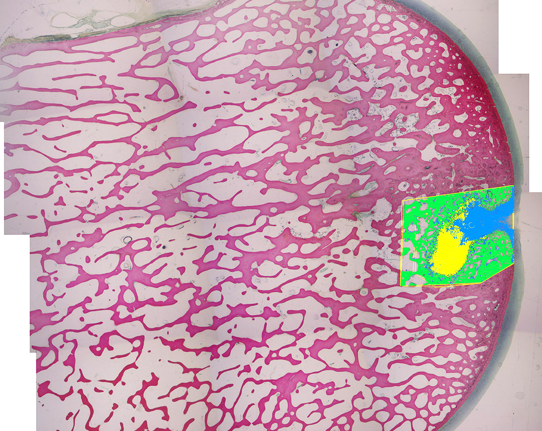
Static Histomorphometry
Static histomorphometery gives a quantitative measurement to what is a happening at a per-determined single time point. Common measurements include:
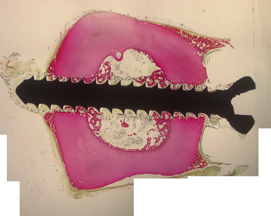
- Areal measurements of bone, fibrous tissue, graft material and implant within a region of interest (ROI)
- Total surface area and percent of implant in contact with bone or fibrous tissue
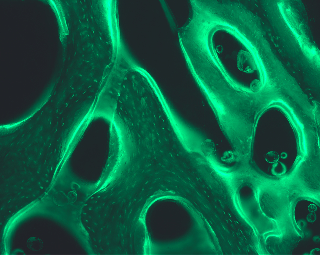
Dynamic Histomorphometry
Dynamic histomorphometery gives a quantitative measurement of what is happening over a pre-determined span of time. This is achieved using fluorescent bone markers which are administered in vivo at specific time points. Common measurements include:
- Mineralized surface
- Mineral apposition rate
- Bone formation rate

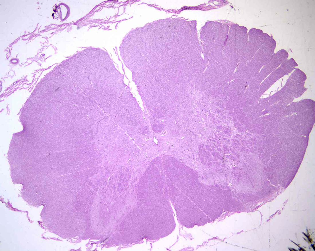
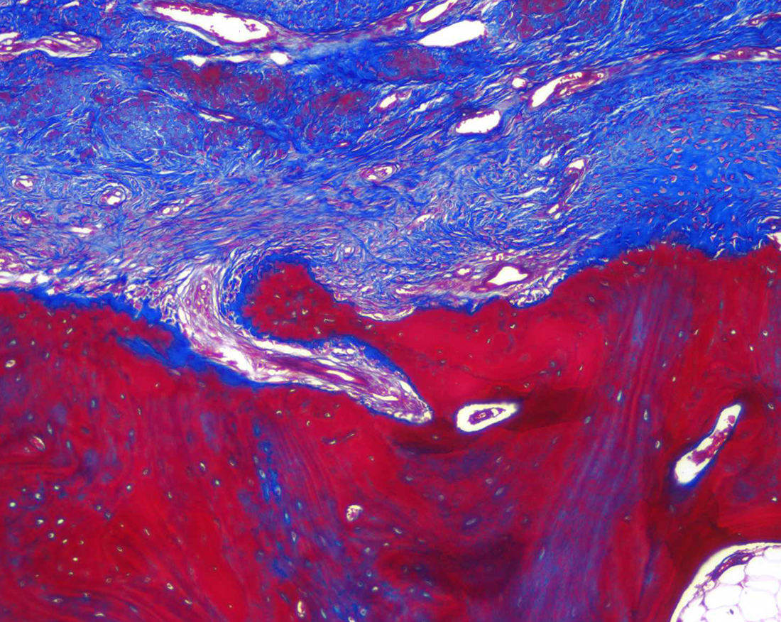
Paraffin Histology
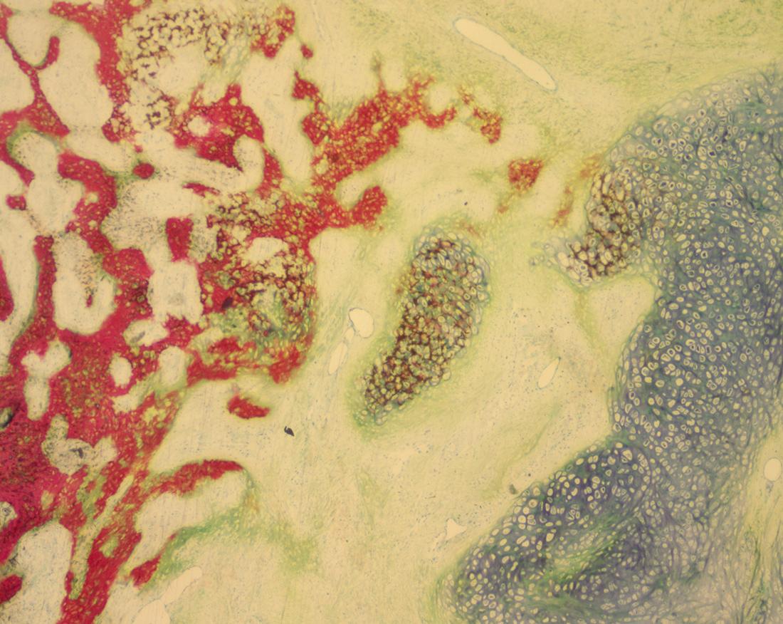
Paraffin techniques are used for soft tissue and decalcified bone samples, producing slides which are appropriate for H&E, special stains, and IHC. Stains routinely used at the OBRL include trichrome, picrosirius red, and safranin O/fast green.
Cryotomy
Frozen tissue sectioning is optimal for some types of IHC or for preserving fats or enzymes which may be lost during paraffin processing.
Immunohistochemistry
Normally completed on paraffin embedded samples, but some IHC tests can be completed on frozen or plastic sections. The most common IHC methods in the OBRL are for visualizing collagen types I and II.
Histology Equipment and Resources
- Exakt cutting and grinding system for plastic ground sections
- Band saw with diamond blades for cutting gross tissue
- Leica RM2255 rotary microtome
- Tissue processor and embedding centers
- ImagePro Premier image capture and analysis software